Xuesong Yuan1 ![]() ,
Xiaoxing Bian1,
Wenfeng Wei1,
Yin Tang2,
Qing Bao1
,
Xiaoxing Bian1,
Wenfeng Wei1,
Yin Tang2,
Qing Bao1
For correspondence:- Xuesong Yuan Email: yuan_cedar@sina.com Tel:+8613775093132
Received: 1 October 2015 Accepted: 7 March 2016 Published: 30 April 2016
Citation:
Yuan X, Bian X, Wei W, Tang Y, Bao Q.
Effect of recombinant human erythropoietin ex
© 2016 The authors.
This is an Open Access article that uses a funding model which does not charge readers or their institutions for access and distributed under the terms of the Creative Commons Attribution License (http://creativecommons.org/licenses/by/4.0) and the Budapest Open Access Initiative (http://www.budapestopenaccessinitiative.org/read), which permit unrestricted use, distribution, and reproduction in any medium, provided the original work is properly credited..
Purpose: To explore the effect of recombinant human erythropoietin (r-HuEPO) on apoptosis in rats after traumatic brain injury.
Methods: A total of 48 traumatic brain-injured Sprague Dawley (SD) rats were obtained by improved Feeney’s traumatic brain injury model, and were randomly divided into four groups: normal saline-treated rats (control) and rats treated with r-HuEPO at doses of 1000 U/kg, 3000 U/kg and 5000 U/kg. Brain tissues were collected on the 7th day after trauma surgery. Apoptotic cells, and NF-kappa B (NF-ĸB)-, c-myc-, and Fas/Fasl-positive cells were identified in brain tissues by immunohistochemical assay.
Results: After treatment with r-HuEPO (3000 and 5000 U/kg), ex
Conclusion: Thus, r-HuEPO may be beneficial for treating traumatic brain injury via inhibition of NF-kappa B and Fas/Fasl ex
Introduction
Traumatic brain injury (TBI) is a major cause of death and persistent disability [1]. In addition, it is reported that over 5 % of patients with moderate injury, and 16 % of patients with severe injury would develop posttraumatic epilepsy [2,3]. The mechanisms underlying secondary brain damage following TBI are complex and involve two main stages: traumatic cerebral edema and delayed neuronal damage [1-3]. Delayed neuronal damage leads to irreversible necrosis or apoptosis of neurons, which could affect the long-term prognosis and quality of life in patients [1,2,4,5]. It is known that NF-kappa B (NF-κB) can result in neuronal damage by apoptosis. In addition, the c-myc gene could also promote apoptosis of neurons. Fatty acid synthase (Fas) is a transmembrane protein belonging to the tumor necrosis factor (TNF) superfamily. Interaction between Fas and its ligand (Fasl) is one of the main pathways for inducing apoptosis [6-9].
Erythropoietin (EPO), which is mainly produced by the kidneys and liver, can affect hematopoietic stem cells present in the bone marrow. Recent research has indicated that EPO can be secreted by astrocytes (paracrine secretion) in the central nervous system (CNS) [10]. Furthermore, the secreted EPO could bind to the EPO receptors present on neuronal membranes nearby. Furthermore, EPO could also protect the nerves from secondary brain damage after TBI by relieving cerebrum edema [11], facilitating neuronal regeneration [12], lowering toxicity of excitatory amino acid neurotransmitters [13], and playing anti-oxidative effect [14]. Our present study used the r-HuEPO to treat traumatic brain injury in rats, and explore the effects of r-HuEPO on the expressions of NF-kB, c-myc and Fas/Fasl in brain tissue.
Methods
Materials
The r-HuEPO was purchased from the Shenyang Shansheng pharmaceutical Co., LTD (Shenyang, China); the primary antibodies of NF-kB and Fas/Fasl were purchased from the Santa Cruz Biotech. (Shanghai, China); the primary antibodies of c-myc were purchased by ZSGB Biotech (Shanghai, China); the secondary antibodies reagents of Envision were purchased from the Fuzhou Maixin Co., Ltd (Fuzhou, China). All the other chemical reagents used in our study were of analytical grade.
TBI rat preparation and grouping
A total of 48 healthy adult male SD rats (weighing between 280 and 320 g) were randomly divided into 4 groups: normal saline treated groups (control) and rhEPO treated groups at the doses of 1000 U/kg, 3000 U/kg rhEPO and 5000 U/kg. TBI rats were prepared by using the Modified Feeney's free-falling model [15]. The trauma preparation device consisted of steel weights falling freely by gravity from a designated height through a Plexiglas tube. The rats were anesthetized with 0.35 % pentobarbital sodium solution (30 mg/kg, intraperitoneal injection (ip). After exposing the left parietal bone by a midline incision, a port with 3 mm diameter was formed by dentistry auger. Thereafter, a 20 g weight was dropped on the exposed dura from 40 cm height. Drugs were administered immediately (ip) after trauma, and the drug was injected every 24 h until 7th day after trauma. All animal experiments were strictly accorded to the international ethical guidelines and the National Institutes of Health Guide concerning the Care and Use of Laboratory Animals, and the experiments were carried out followed the approval of the Animal Experimentation Ethics Committee of our Hospital (Ethical approval No.201312-A87).
Sample collection and evaluation
Brain tissues were collected at 7th day after trauma and fixed in formaldehyde. The damaged area of brain was imbedded with paraffin, and then, sliced. The NF-κB-, c-myc- and Fas/Fasl- positive cells, as well as apoptotic cells in brain tissues were detected by immuno-histochemistry assay. Cells with yellowish-brown grains in cell nucleus or endochylema were considered as positive expressions.
Statistical analysis
All data are expressed as mean ± standard deviation (SD) and analyzed by SPSS 10.0 statistical analysis software using analysis of variance (ANOVA). Differences were considered significant at p < 0.05.
Results
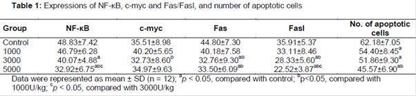
As shown in and Plate 1, the positive expression levels of NF-κB and Fas/Fasl in r-HuEPO treated TBI rats at the doses of 3000 and 5000 U/kg were significantly decreased (p < 0.05), compared with the normal saline treatment group (control group), particularly in the 5000 U/kg treated group (p < 0.01). However, no significant difference in the c-myc expression was observed in each r-HuEPO treatment group (p > 0.05). Compared with the 1000 U/kg r-HuEPO treated group, the positive expressions of c-myc and Fas/Fasl were significantly lower in 3000 U/kg r-HuEPO group (p < 0.05), and the positive expression levels of NF-κB and Fas/Fasl in 5000 U/kg r-HuEPO treated group were significantly lower (p < 0.05). Compared with the 3000 U/kg r-HuEPO treated group, only the positive expression levels of NF-κB and Fasl in 5000 U/kg r-HuEPO group were significantly lower (p < 0.05). The results also indicated that the number of apoptotic cells in r-HuEPO treated group (5000 U/kg) was significantly lower compared with the control group (p < 0.05, Plate 2).
Discussion
After traumatic brain injury, the overexpression of pro-apoptotic genes will increase the tardus injury and irreversible degeneration necrosis or apoptosis of neurons, causing more serious consequences than traumatic brain edema. In this study, after traumatic brain injury, a large number of apoptotic cells were detected in the cerebral cortex of the control rats, indicating that neuronal apoptosis plays a curial role in secondary brain damage after traumatic brain injury. Interestingly, after treating with r-HuEPO, the expressions of pro-apoptotic proteins including NF-κB, c-myc and Fas/Fasl were significantly decreased compared with control group. In particular, after administration of r-HuEPO at the dose of 5000 U/kg, the expressions of pro-apoptotic genes were suppressed most obviously. However, no obvious difference was observed in the r-HuEPO treatment rats for the expression of c-myc gene. The findings demonstrate that r-HuEPO has treatment effects on the expression of NF-κB, c-myc, Fas and Fasl. In addition, our results also indicate that large dose of r-HuEPO has a strong suppressive effect on NF-κB and Fasl expression.
Clinically reports revealed that prolonged usage of large doses of EPO may result in lots of side effects, such as increased blood volume, increased blood viscosity, and high blood pressure due to the proliferation of red blood cells [15,16]. Recently, Erbayraktar et al have developed a novel kind of EPO that does not contain the sialic acid group [17]. Interestingly, Erbayraktar found that this EPO did not promote the proliferation of red blood cells, and that it had a short half-life in the blood plasma, thereby avoiding the above-mentioned side effects. It also performed a better nerve-protective effect in the nervous system through the blood-brain barrier. Combined with the results of our study, it can be assumed that EPO might be used in clinical treatments to reduce neuronal damages after traumatic brain injuries.
Conclusion
The findings of this investigation suggest that EPO effectively suppresses the pro-apoptotic proteins, NF-κB and Fas/Fasl, after traumatic brain injuries in rats, and hence reduce neuronal damage. These findings provide some experimental evidence for the clinical use of EPO in treating severe traumatic brain injuries, especially in improving long-term prognosis and improving patients’ quality of life.
References
Archives


News Updates